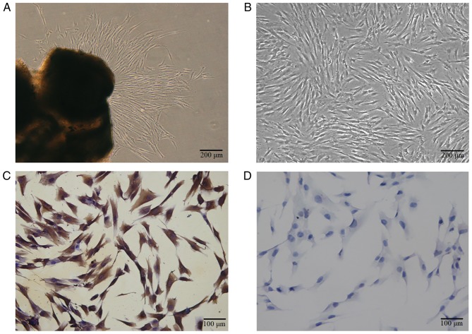
Figure 2.
Characterization and identification of hPDLCs (A) Primary cells grew out from tissue explants. (B) The primary hPDLCs exhibited a spindle shape. (C) Immunocytochemistry staining was positive for vimentin. (D) Immunocytochemistry staining was negative for cytokeratin. For (A) and (B): Scale bar, 200 µm. For (C) and (D): Scale bar, 100 µm. hPDLCs, primary human periodontal ligament cells.

