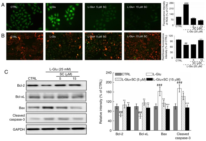
Figure 2.
SC reduces ROS accumulation, restores MMP dissipation, and regulates anti- and pro-apoptotic protein expression. HT22 cells were pre-incubated with 5 and 15 µM SC for 3 h, and exposed to L-Glu for another 12 h. (A) Representative immunofluorescent images of HT22 stained with 2,7-dichlorofluorescein diacetate to visualize intracellular ROS (magnification, ×200; n=6). SC suppressed the accumulation of ROS, as demonstrated by reductions in green fluorescence intensity. (B) Immunofluorescence staining with JC-1 revealed that SC restored the dissipation of MMP (magnification, ×200; n=6). The quantitative data of the ratio of red to green fluorescence intensity is presented on the right. (C) Following treatment with SC, cells were treated with L-Glu for another 24 h. Western blot analysis demonstrated that 15 µM SC was able to restore the expression of anti-apoptotic (Bcl-2 and Bcl-xL) and pro-apoptotic (Bax and cleaved caspase-3) proteins to normal levels. Quantification data were normalized to GAPDH and expressed as a percentage of CTRL (n=3). ##P<0.01, ###P<0.001 vs. CTRL; *P<0.05, **P<0.01, ***P<0.001 vs. L-Glu. Bax, Bcl-2-associated X protein; Bcl-2, B-cell lymphoma 2; Bcl-xL, B-cell lymphoma-extra large; CTRL, control; L-Glu, L-glutamic acid; MMP, mitochondrial membrane potential; ROS, reactive oxygen species; SC, scutellarin.

