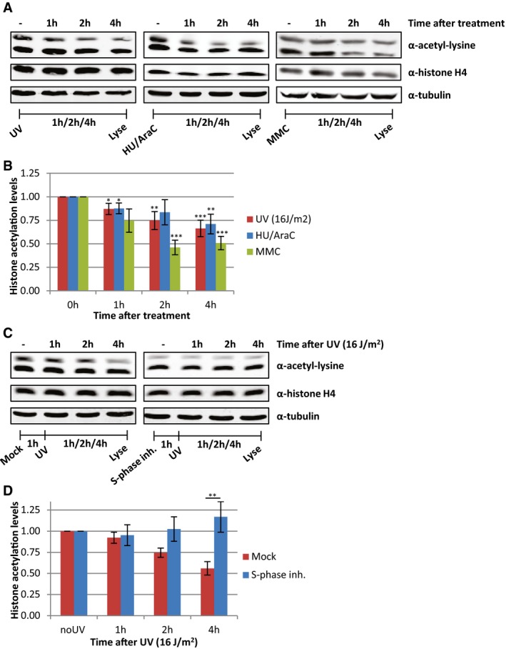
Representative Western blots of histone acetylation levels of HeLa cells obtained at the indicated time points after UV irradiation (16 J/m2), HU/AraC treatment (100 mM/10 μM), or MMC treatment (10 μg/ml). Blots were stained with α‐acetyl‐lysine (top panel), α‐histone H4 (middle panel), and α‐tubulin (bottom panel).
Quantification of average histone acetylation signal, normalized to loading control. N ≥ 3. Error bars represent SEM. Significant differences, calculated with t‐test, between treated and mock conditions are indicated with *P < 0.1, **P < 0.05, and ***P < 0.01.
Representative Western blots, stained with the indicated antibodies of HeLa cells pre‐treated o/n with an S‐phase inhibitor (PHA 767491 hydrochloride, 10 μM) and lysed at indicated time points after UV (16 J/m2).
Quantification of the α‐acetyl‐lysine signals, normalized against histone H4 levels, N = 5, error bars represent SEM. Significant differences, calculated with t‐test, between UV‐irradiated and inhibitor‐treated conditions are indicated with **P < 0.05.