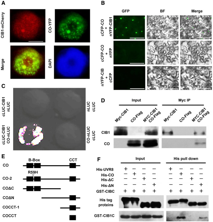
Fluorescent microscopy images showing that CIB1 co‐localizes with CO in nuclear speckles. Scale bars: 2 μm.
BiFC assays to show in vivo protein interactions. Epidermal cells of Nicotiana benthamiana leaves were co‐transformed with cCFP‐CO and nYFP‐CIB1, with cCFP‐CO and nYFP, or with cCFP and nYFP‐CIB1. BF, bright field. Merge, overlay of the YFP and bright field images. Scale bars: 50 μm.
Split‐LUC assay showing that CIB1 interacts with CO. Leaf epidermal cells of N. benthamiana were co‐transformed with CO‐nLUC and cLUC‐CIB1 or cLUC or nLUC with cLUC‐CIB or cLUC.
Co‐IP assays of samples prepared from 10‐day‐old 35S:Myc‐CIB1, 35S:CO‐Flag, or 35S:Myc‐CIB1/35S:CO‐Flag seedlings grown in LDs.
Schematic representation of CO used in this work was showing. CO contains two B‐Box domains and a CCT domain.
In vitro pull‐down assays showing the interaction between CIBC and CO, or CIBC and COΔN and the lack of interaction between CIB1C and COΔC. His‐TF tagged CO, COΔN, COΔC, or His‐TF tagged UVR8 was mixed with GST‐CIB1C purified from Escherichia coli, and His antibody was used for the in vitro pull‐down assay. The products were analyzed by immunoblots probed with the anti‐CIB1C antibody or the anti‐His antibody (UVR8, CO, COΔC, COΔN).
Data information: For each experiment, two or more biological replicates were performed.

