-
A
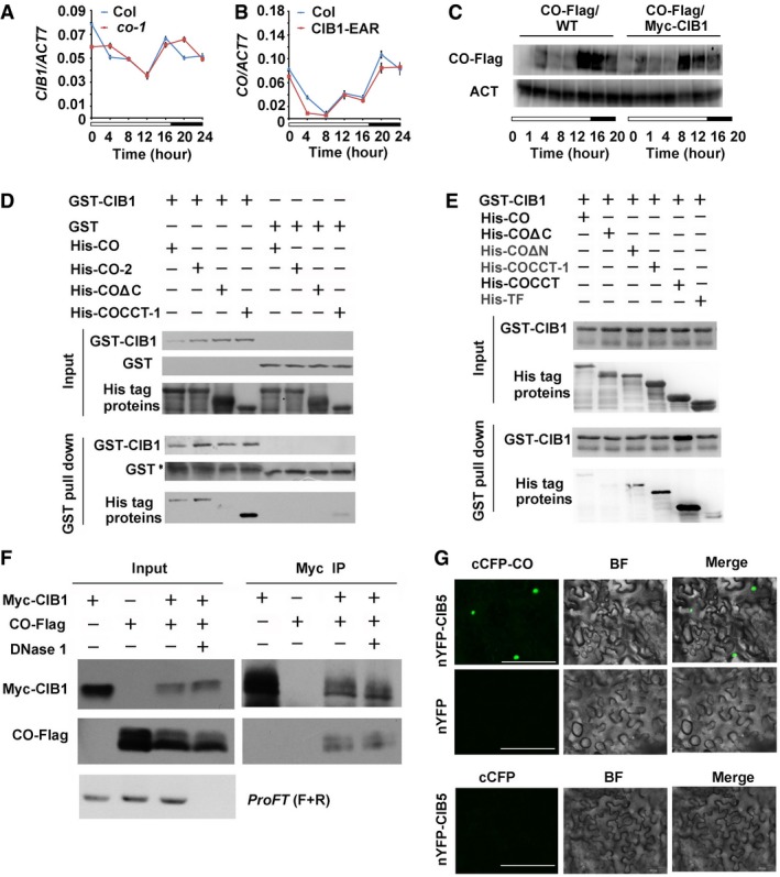
qPCR results showing the mRNA expression of CIB1 in the WT and the co‐1. Error bars represent SD of three technical replicates of qPCR. The experiment was performed at least three times with similar results.
-
B
qPCR results showing the mRNA expression of CO in the WT and CIB1‐EAR transgenic lines. Error bars represent SD of three technical replicates of qPCR. The experiment was performed at least three times with similar results.
-
C
Immunoblots showing CO protein levels in transgenic lines expressing 35S:CO‐Flag and 35S:CO‐Flag/35S:Myc‐CIB1 over the course of 20 h under LDs. Samples were separated on 10% SDS–PAGE, blotted, probed with the anti‐Flag antibody, stripped and re‐probed with the anti‐actin antibody (ACT).
-
D, E
In vitro pull‐down assays showing the interaction between CIB1 and CO, or CIB1 and COΔN, or CIB1 and COCCT‐1, or CIB1 and COCCT and the lack of interaction between CIB1 and COΔC. His‐TF tagged CO, CO‐2, COΔN, COΔC, COCCT‐1, or COCCT was mixed with GST‐CIB1 purified from Escherichia coli, and TF antibody was used for the in vitro pull‐down assay. The products were analyzed by immunoblots probed with the anti‐GST (CIB1) or the anti‐TF antibody (CO, CO‐2, COΔN, COΔC, COCCT‐1, COCCT). *Indicates non‐specific bands.
-
F
Co‐IP assays showing in planta interactions between CO and CIB1 with or without FT DNA. Protein extracts from transgenic lines co‐expressing Myc‐CIB1 and CO‐Flag were treated with or without DNase I at 22°C for 15 min before used in the co‐IP assay. ProFT (F+R): 1,000 bp FT promoter was amplified using PCR to verify the digestion of the DNase I.
-
G
BiFC assays of the in vivo protein interaction. Epidermal cells of Nicotiana benthamiana leaf were co‐transformated with cCFP‐CO and nYFP‐CIB5, with cCFP‐CO and nYFP. BF, bright field. Merge, overlay of the YFP and bright field images. Scale bars: 50 μm.

