. 2018 Sep 28;51(3):150–157. doi: 10.5115/acb.2018.51.3.150

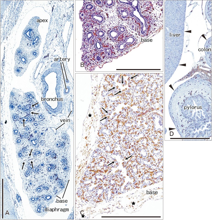
Fig. 3. CD34 immunohistochemistry of the lung and pleura. Sagittal sections. (A) The left lung from a specimen at 9 weeks (pseudoglandular phase; 39 mm crown-rump length [CRL]). (B) The left lung from a specimen at 15 weeks (a mixture of the pseudoglandular and canalicular phases; 115 mm CRL). (C) The left lung from a specimen at 30 weeks (saccular phase; 250 mm CRL). Panels B and D are photos of the same section and panel D displays the liver, pylorus and transverse colon. CD34 immunoreactivity is restricted in the developing vessels (arrows in A). At 15 weeks, the subpleural tissue is thin at the lung base (B), while no or little serosal tissue is seen along the liver, pylorus and colon (arrowheads in D). At 30 weeks, the subpleural tissue contains multilayered fibers expressing CD34 weakly (C). Capillaries adjacent to alveoli express CD34 strongly (arrows in C). Scale bars=1 mm.

Fig. 3