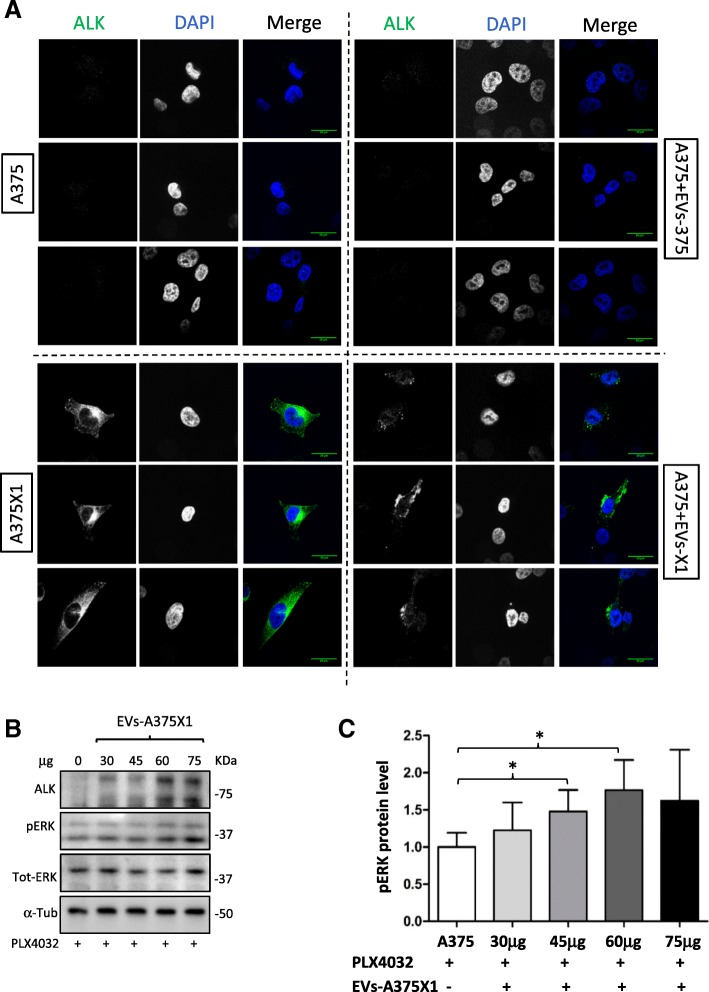
Fig. 6.
Functional ALKRES is transferred to sensitive cells via EVs. (a) Sensitive A375 melanoma cells were co-cultured with 10 μg of both EV-A375 and EV-A375X1. After 24 h, untreated A375 cells, resistant A375X1 cells and A375 co-cultured with both types of EVs were fixed and stained for ALK. Images were captured by fluorescence confocal microscopy. Representative images of two biological replicates. Scale bar, 20 μm. Blue: nucleus; green: ALK. (b) Sensitive A375 cells were treated with 1 μM of PLX4032. After 1 h, increasing concentrations of resistant EVs were added to the cells for additional 6 h. α-Tubulin was used as a loading control; representative blots of three biological replicates are shown. (c) Quantification of pERK levels, normalised to the untreated control. Error bars represent the standard deviation of three biological replicates. Statistical significance was determined using paired Student’s t-tests. *p < 0.05, **p < 0.01, ***p < 0.001

