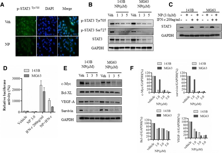
Fig. 3.
NP blocks STAT3 activation and STAT3-dependent gene expression. a, MG63 cells treated with vehicle or 3 μM NP for 24 h were analyzed by immunofluorescence using anti-phospho-STAT3Tyr705 antibodies. b, Cytoplasmic extracts from osteosarcoma cells treated with NP were analyzed by Western blot using anti-STAT3, anti-phospho-STAT3Tyr705, anti-phospho-STAT3Ser727, and anti-GAPDH antibodies as described in the Methods section. c, Cytoplasmic extracts prepared from143B and MG63 cells treated with vehicle, NP, and IFN-γ for 24 h were analyzed by Western blot analysis. d, 143B and MG63 cells transiently transfected with GAS-luciferase reporter plasmids were treated with vehicle and NP (3.0 μM) for 24 h, and luciferase activity was analyzed. e, 143B and MG63 cells were treated with vehicle or indicated concentrations of NP for 24 h. The cytoplasmic extracts prepared following the treatment were analyzed by Western blot using anti–VEGF-A, anti–c-Myc, Bcl-2, anti–Bcl-xl, anti-survivin, and anti-GAPDH antibodies. f, Quantitation of Western blot using densitometry. *P < 0.05 versus vehicle control; **P < 0.01 versus vehicle control

