Fig. 2.

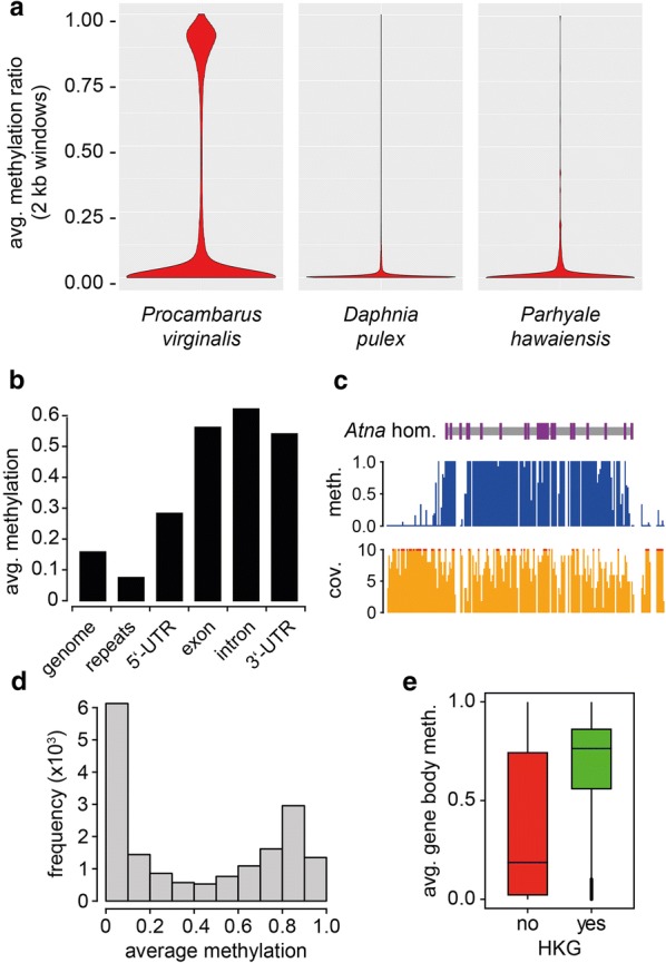
Characterization of the marbled crayfish methylome. a Comparative analysis of known crustacean methylomes. Violin plots show average CpG methylation levels of 2-kb sliding windows. b Methylation levels of the genome and of predicted gene features. c Representative Genome Browser track for a methylated gene, showing methylation ratio (blue) and coverage (orange). Red dots denote coverages > 10 ×. d Histogram showing the frequencies of average gene body methylation levels in bins of 0.1. e Boxplots showing the distribution of methylation ratios for non-housekeeping genes (red) compared with housekeeping genes (green)
