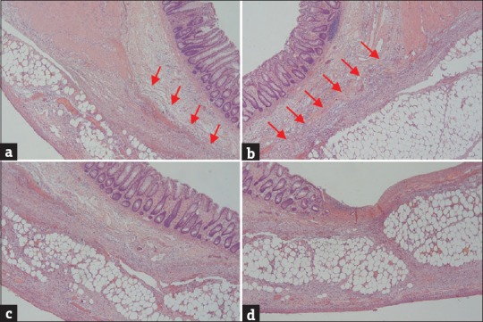
Figure 3.

(a and b) The affected colon showed the full-thickness absence of muscularis propria (arrow) with a blunt-end appearance in the absence of necrosis, significant inflammatory cell infiltrates, or granulation tissue. (c) The mucosa, muscularis mucosae, submucosal layers, and serosal layers were normal in the absence of muscularis propria. (d) The mucosa showed focal ulceration with sloughing epithelium, necrotic debris, and aggregated neutrophils. The muscularis mucosae was normally preserved in the area of the muscular defect. The submucosal and subserosal layers revealed vascular congestion, mild inflammation, and adipose tissue replacement in the colonic wall (H and E, ×40)
