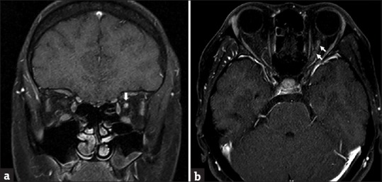
Figure 2.

(a) The presence of a T1 high signal with contrast enhancement over the posterior segment of the optic nerve in the coronal view on magnetic resonance imaging (arrow). (b) The presence of a T1 high signal from the retrobulbar to the intracanalicular segment of the left optic nerve (arrow), compatible with acute optic neuritis
