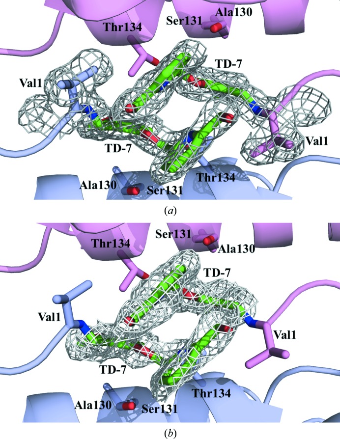
Figure 5.
Close-up view of TD-7 (green sticks) bound at the α-cleft of R2 Hb with contoured electron density. Hb subunits are shown as ribbons (α1 subunit in pink, α2 subunit in purple). Note how the pyridine rings are engaged in a π–π stacking interaction to stabilize the binding and the R-state Hb (see text). For clarity, not all binding-site residues are shown but are described in the text. (a) Final 2F o − F c refined electron-density map contoured at 1.0σ. (b) Initial F o − F c electron-density map (prior to addition of TD-7 to the model) contoured at 3.0σ.

