Figure 2.
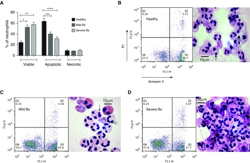
Blood neutrophils from patients with mild and severe bronchiectasis survived longer and underwent less apoptosis when compared with healthy volunteers. Blood neutrophils from patients with mild and severe bronchiectasis in a stable state and healthy volunteers were cultured for 20 hours and cell viability (AnnV−/PI−), apoptosis (AnnV+/PI−), and necrosis (AnnV+/PI+) were assessed by flow cytometry. (A) n = 8 in each group; percentage of viable, apoptotic, and necrotic neutrophils in each group; *P < 0.05; **P < 0.01; ***P < 0.001; ****P < 0.0001. One-way ANOVA with Bonferroni correction for multiple comparisons used for all three groups compared; comparing mild and severe bronchiectasis with healthy control subjects in viable, apoptotic, and necrotic neutrophils. (B–D) Representative flow cytometry plots and cytocentrifuge preparations at 20 hours. AnnV = annexin V; Bx = bronchiectasis; FL1-H and FL2-H = fluorescence indices–height; PI = propidium iodide. Black arrow = dark pyknotic apoptotic nucleus; gray arrow = multilobulated viable nucleus.

