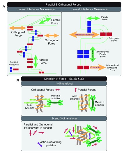
Figure 2. Direction of forces at the lateral cell–cell adhesion interface.
( A) Parallel and orthogonal forces are shown at the macroscopic multi-cellular (left panel) and mesoscopic cellular (right panel) scales. Cell–cell adhesions at the tip of a lateral membrane protrusion experience orthogonal force, whereas cell–cell adhesions at the trunk of the same membrane protrusion experience parallel force (right panel). ( B) Forces in one, two, and three dimensions can be generated through spatial organization of actin dynamics and actomyosin activities. Orthogonal and parallel one-dimensional forces can be generated with actin filaments arranged orthogonal and parallel to the plasma membrane, respectively (upper panel). Orthogonal and parallel two- and three-dimensional forces can be generated with a cross-linked actin network to support processes on the lateral cell–cell adhesion interface (lower panel).

