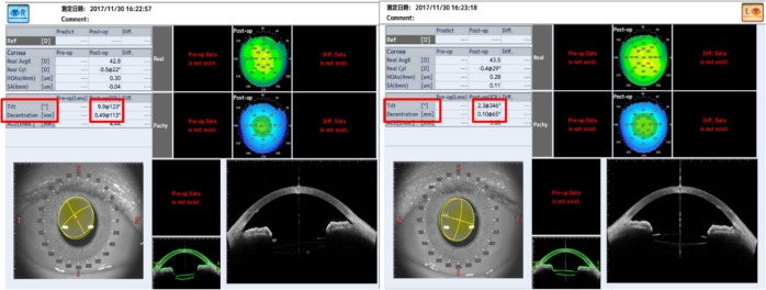
Figure 8.
Image of the anterior segment OCT.
Notes: R: right eye with the single-piece trifocal IOL in the bag. L: left eye with the three-piece bifocal IOL in the sulcus of the optic capture. Degree of tilt and decentration is indicated by the red box.
Abbreviations: IOL, intraocular lens; OCT, optical coherence tomography.

