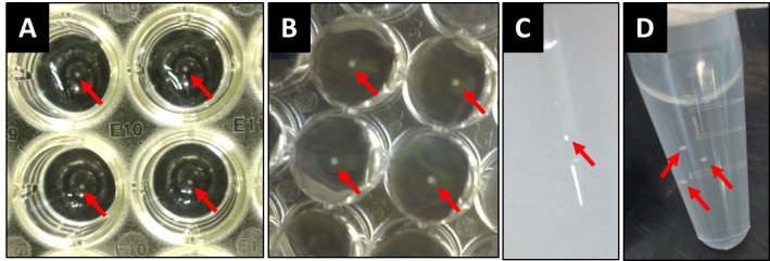
Figure 2. Working with spheroids.

Spheroids can be seen in the wells from above (A) and below (B) the plate (5 days spheroids in the picture, 20,000 cells). Spheroids can be easily aspirated with a pipette tip (C) and transferred into centrifuge tubes (D). Spheroids are indicated with arrows. As a reference, the wells from (A) and (B) are from a 96-well plate.
