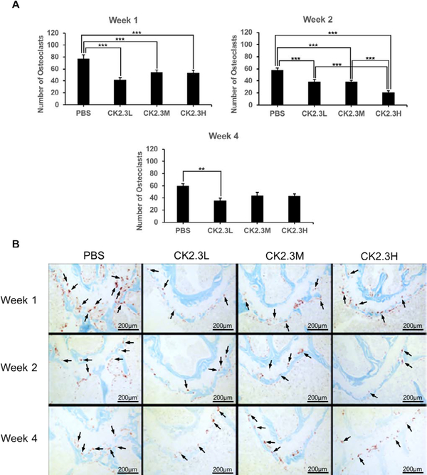
Figure 4:
CK2.3 suppressed osteoclastogenesis in vivo. Images of distal growth plate region of femur were taken. Osteoclasts were stained with TRAP staining assay and appeared as purple. Osteoclast number was significantly decreased one week (p-value<0.0001) and two weeks (p-value<0.0001) after the first injection at all concentrations. But only CK2.3L suppressed osteoclastogenesis four weeks (p-value<0.01) after the first injection. A) quantify number of osteoclasts in each treatment at different time points. B) representative images of each treatment at different time points. Five to six mice were used per treatment group at each time point. Arrows point to representative osteoclasts (not all osteoclasts were pointed out). Statistically significance was performed by ANOVA followed by Tukey-Kramer (** and ***=significant, p-value<0.01 and p-value<0.0001, respectively).

