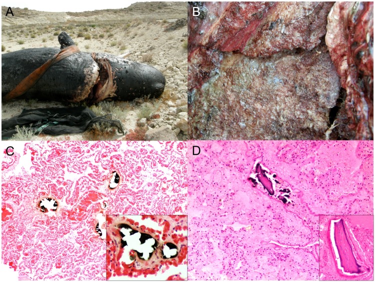
Fig 7. Panel of vessel collision-associated pathologic findings in cetaceans stranded in the Canary Islands (2006–2012).
A) Cervico-occipital fracture (animal no. 142; Physeter macrocephalus). There is a single, well-demarcated, deep incisive cut through the dorsal occipital region and the atlas vertebra. B) Cervico-occipital fracture (animal no. 142; P. macrocephalus). Multiple bone fracture surfaces are stained with blue antifouling paint from the vessel cutting edges (presumably the keel). C) Fat embolism (animal no. 86; Kogia breviceps). The microvasculature of the lung parenchyma, mainly alveolar capillaries, contains multiple osmium-tetroxyde-positive (black) fat emboli. OsO₄ (postfixation technique) and H&E. Inset (animal no. 86; K. breviceps): fat embolus obliterates and expands the vascular lumen. OsO₄ (postfixation technique) and H&E. D) Pulmonary bone emboli (animal no. 60; K. breviceps). There are multiple osseous emboli in the pulmonary microvasculature. H&E. Inset: Embolic osseous fragments consists of lamellar (mature) bone with multiple presumably viable osteocytes in lacunae. H&E.

