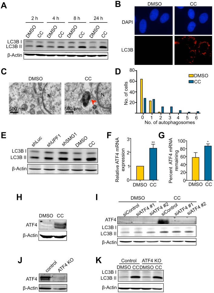
Fig 5. CC induces autophagy independently of the expression and stabilization of the NMD target ATF4.
A. Effects of CC on LC3BII levels in U2OS cells. Cells were treated with DMSO or CC (10 μM) for 24 hours and collected at the indicated time points. B. Immunofluorescence detection of LC3B foci in U2OS cells after 24-hour treatment with DMSO or CC (10 μM). DAPI was used to visualize nuclei. C. Representative electron microscopy images of U2OS cells after 24-hour treatment of with DMSO or CC (10 μM). The arrow marks an electron-dense autophagosome. N, nucleus. D. Quantification of autophagosomes formed after 24-hour treatment with DMSO or CC (10 μM). Two sections were made from 2 different blocks for each sample. Each section is 80 nm thick and 1.75 mm long. The number of autophagosomes per cell from the two sections was counted for each sample. In total, 89 cells were counted in DMSO control, and 88 cells were counted in the CC-treated sample. E. Effects of shRNA-mediated knockdown of UPF1 or SMG1 on LC3BII levels in U2OS cells, compared to the effects in U2OS cells treated with DMSO or CC (10 μM) for 24 hours. F. Effects of CC on ATF4 mRNA expression in U2OS cells treated with DMSO or CC (10 μM) for 24 hours. mRNA expression of DMSO-treated cells was normalized to 1. Data represent the mean ± SD of three independent experiments. **p ≤ 0.01 (paired t-test). G. Effects of CC on ATF4 mRNA stability in U2OS cells. Cells were treated with DMSO or CC (10 μM) for 24 hours followed by treatment with actinomycin D for 6 hours. Total RNA was collected before and after actinomycin D treatment. ATF4 mRNA levels was measured by RT-qPCR. Data represent the mean ± SD of three independent experiments. *p ≤ 0.05 (paired t-test). H. ATF4 protein levels after 24-hour treatment with DMSO or CC (10 μM). I. Effects of siRNA-mediated knockdown of ATF4 on LC3BII levels in U2OS cells treated with DMSO or CC (10 μM) for 24 hours. J. ATF4 protein levels in WT or knockout U2OS cells. Because the basal level of ATF4 is below detection, cells were treated with 0.2 μM Thapsigargin (a known ER stressor that induces ATF4) for 4 hours before ATF4 western blot. K. Effects of ATF4 KO on LC3BII levels in WT or ATF4-KO cells treated with DMSO or CC (10 μM) for 24 hours.

