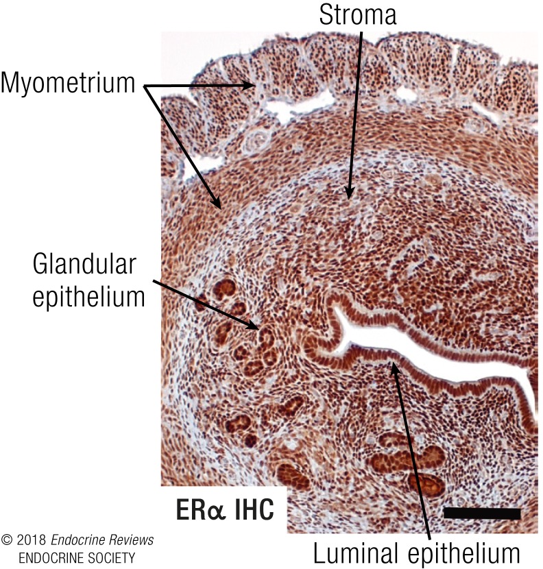
Figure 4.
Mouse uterus as ERα-mediated E2 response model. Cross-section of a mouse uterus stained by immunohistochemistry (IHC) with an antibody to ERα illustrating plentiful ERα in all cells, including the luminal and glandular epithelial cells, stromal cells, and myometrial cells. Photo taken with ×10 objective, scale bar = 100 μM.

