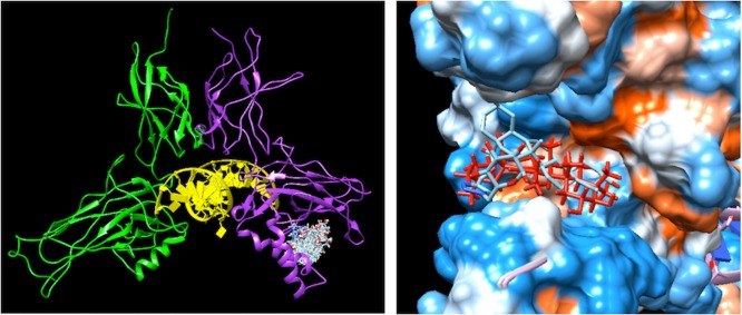
Figure 3.
Left: poses resulting from the docking of compound 18 on p52:v-Rel (PDB accession code 3do7). Subunit v-Rel is shown in green, subunit p52 in purple, and DNA in yellow. Right: alignment of the top score solution of MA (red) and compound 18 (cyan). The protein is shown as a surface colored according to the hydrophobicity on the Kyte–Doolittle scale, ranging from dodger blue, for the most hydrophilic, to white and orange red, for the most hydrophobic.

