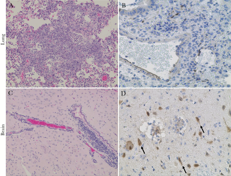
Figure 2.
Pathological lesions and viral antigen distribution in the lung and brain of Syrian hamsters following aerosol exposure to Nipah virus Malaysia strain (NiV-M) strain. Hematoxylin and eosin staining (A, C) and NiV nucleoprotein staining (brown staining) (B, D) of both lung and sagittal brain sections from NiV-M–infected hamsters. Hyperplasia type II pneumocytes with inflammatory cells in alveoli (A), NiV-M–infected endothelial cells from large and medium size pulmonary vessels (B), severe perivascular cuffing by mononuclear cells in meninges (C), and NiV-M–infected neurons (D) (arrows). Initial magnification 20 × (A, C), 40 × (B, D).

