Figure 2.

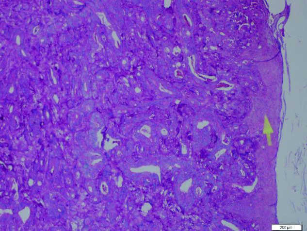
Pathologic section of metastatic foci of the skin: irregular and atypic cells forms abortif adenoidal glands with pseudostratified columnar epithelium; luminal mucus and inflammatory cells. (Hematoxylin and eosin X100)

Pathologic section of metastatic foci of the skin: irregular and atypic cells forms abortif adenoidal glands with pseudostratified columnar epithelium; luminal mucus and inflammatory cells. (Hematoxylin and eosin X100)