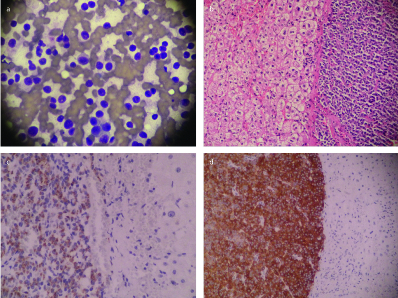
Figure 2.
a–d. Microscopic features of the excised liver mass. (a) Individually localized neoplastic cells with neuroendocrine characteristics (400×, May-Grünwald Giemsa). (b) Liver metastasis of neuroendocrine tumor (200×, Hematoxylin and eosin). (c) Liver tumor with positive synaptophysin staining (200×, immunohistochemistry). (dd) Liver tumor with positive chromogranin A staining (200×, immunohistochemistry)

