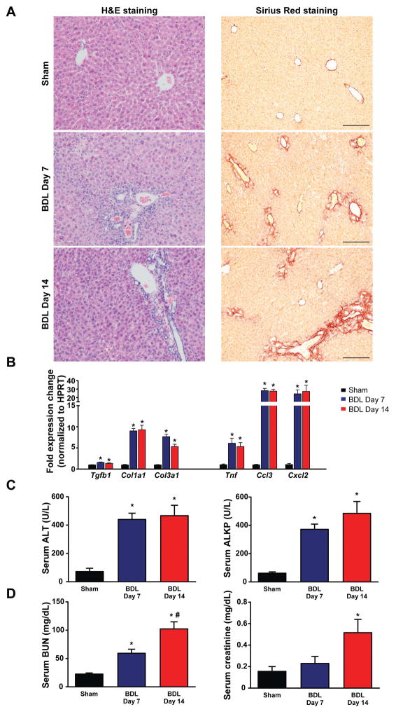
Fig. 1. Time-dependent changes in the markers of liver inflammation and fibrosis in bile duct-ligated mice.
(A) Histological assessment of hepatic necrosis, and fibrosis in BDL mice. Scale bar represent 200 μm. (B) Expression of fibrotic markers TGF-β(Tgfb1), collagen 1(Col1a1), collagen 3(Col3a1) and inflammatory markers TNF-α(Tnf), MIP-1-α(Ccl3), MIP2(Cxcl2), in livers of sham operated mice (Sham) or in mice subjected to BDL and sacrificed on postoperative day 7 (BDL Day 7) or day 14 (BDL Day 14), respectively. (C) Enzymatic markers of liver and (D) kidney injury. Results are mean±S.E.M. *p<0.05 vs. sham #p<0.05 vs. BDL Day 7, n=5–6.

