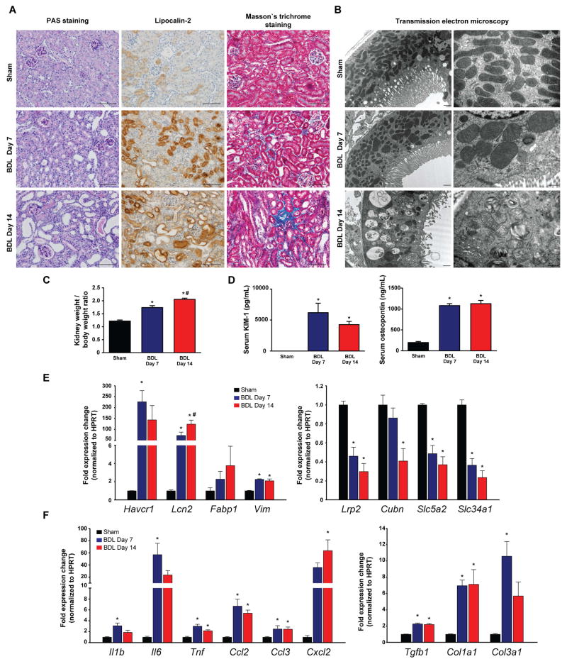
Fig. 2. Bile duct-ligation induces massive tubular damage.
(A) Histological assessment of tubular injury (PAS staining, and lipocalin-2 immunohistochemistry), as well as tubulointerstitial fibrosis (Masson’s trichrome staining). Scale bar represent 100 μm. (B) Transmission electron microscopic images of proximal tubular epithelial cells. Original magnification on left panels are 10,000X; high power fields are 25,000X. (C) Kidney/body weight ratio during the course of study. (D) Serum KIM-1 and osteopontin protein levels of sham operated mice (Sham) or of mice subjected to BDL and sacrificed on postoperative day 7 (BDL Day 7) or day 14 (BDL Day 14), respectively. (E) Expression of tubular injury markers KIM-1 (Havcr1), Lipocalin-2 (Lcn2), fatty acid binding protein 1 (Fabp1) and vimentin (Vim), and genes involved in absorptive function of proximal tubuli megalin (Lrp2), cubilin (Cubn), SGLT2 (Slc5a2), and sodium-dependent phosphate transport protein 2A (Slc34a2) in kidneys of sham operated mice (Sham) or mice subjected to BDL and sacrificed on postoperative day 7 (BDL Day 7) or day 14 (BDL Day 14), respectively. (F) Expression of inflammatory markers IL-1β(Il1b), IL-6(Il6), TNF-α(Tnf), MCP-1(Ccl2), MIP-1-α(Ccl3), MIP2(Cxcl2), and expression of fibrotic markers TGF-β(Tgfb1), collagen 1(Col1a1), collagen 3(Col3a1), in kidneys of sham operated mice (Sham) or in mice subjected to BDL and sacrificed on postoperative day 7 (BDL Day 7) or day 14 (BDL Day 14), respectively. Results are mean±S.E.M. *p<0.05 vs. sham, n=6.

