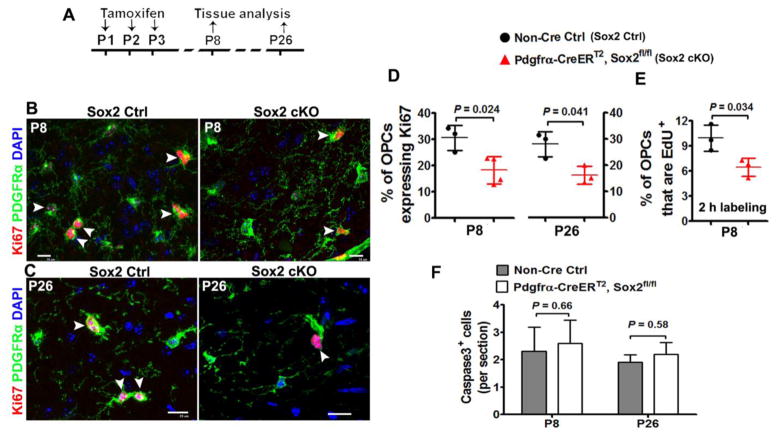
Figure 7. Sox2 regulates OPC proliferation in the postnatal mouse spinal cord.
A, transgenic mice and experimental designs for panels B–E. Thymidine analog EdU was injected intraperitoneally into Sox2 Ctrl and cKO mice 2 h before tissue harvesting. B–C, representative confocal images showing reduction of Ki67+PDGFRα+ cycling OPCs in the white matter of spinal cord at P8 (B) and P26(C). Arrowheads point to Ki67+PDGFRα+ proliferating cells. Scale bars = 10 μm. D, percentage of PDGFRα+ OPCs that are Ki67-positive in the spinal cord white matter at P8 and P26. Two-tailed Student’s t test, t(5) = 3.198 P8, t(4) = 3456 P26. E, percentage of PDGFRα+ OPCs that are EdU-positive (2 h pulse labeling) in the spinal cord white matter at P8. Two-tailed Student’s t test, t(4) = 3.175. F, active Caspase 3+ cells per section. Two-tailed Student’s t test, t(6) = 0.626 P8, t(4) = 0.702 P26.

