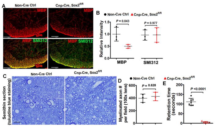
Figure 9. myelination assessment in the spinal cord of Cnp-Cre:Sox2fl/fl mutants at P8 and P60.
A, immunohistochemistry of MBP and axonal marker SMI312 in the ventral medial white matter at P8, scale bar = 100μm. Note the SMI312+ axons are highly myelinated in the ventral medial white matter of spinal cord at P8, thus rendering the overlapped images yellow. B, quantification of the relative intensity of MBP and SMI312 in the ventral medial white matter at P8. Two-tailed Student’s t test, t(4) = 2.922 MBP, t(4) = 0.030 SMI312. C–D, representative images of toluidine blue staining of myelin on semithin (500 nm) sections in the corticospinal tract at P60 (C, scale bar=10μm) and quantification of myelination axon numbers (D). Two-tailed Student’s t test, t(4) = 0.524. E, retention time on the accelerating Rotarod of Cnp-Cre:Sox2fl/fl mutant and control mice at P60. Two-tailed Student’s t test with Welch’s correction, t(6) = 14.85.

