
Figure 3.
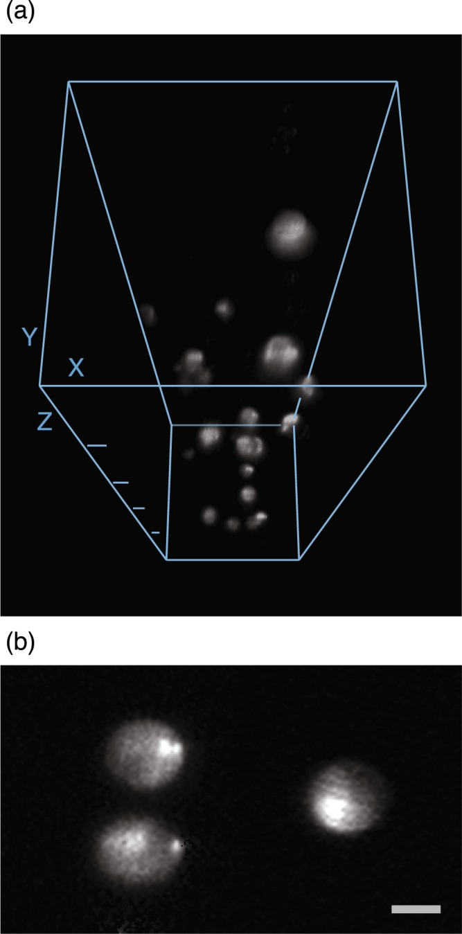
Three-dimensional reconstruction (a) and an optical section (b) of fluorescent microspheres embedded in agarose gel. We used the 3D Viewer of ImageJ to reconstruct the three-dimensional view from 332 sequential optical sections with a 3-μm depth interval. For each optical section, we integrated four images of 1-ms exposure obtained by translating the fiber bundle to four different positions, equivalent to an overall frame rate of 250 fps. The depth difference between adjacent Z ticks in (a) is 200 μm, and the scale bar in (b) is 10 μm. The microspheres are 15 μm in diameter.