Fig. 2.
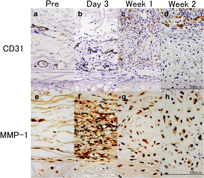
Endothelial (CD31-positive) cells and MMP-1-positive cells. a–d: Biochemical evaluation of rabbit perichondrium tissue immunostained with anti-CD31 peroxidase-conjugated antibody, e–h: biochemical evaluation of rabbit perichondrium tissue immunostained with anti-MMP-1 peroxidase-conjugated antibody. a, e: Pre-injection of bFGF; b, f: day 3 post bFGF-injection; c, g: week 1 post bFGF-injection; d, h: week 2 post bFGF-injection. a CD31-positive cells were scarcely observed around the perichondrium layer in the noninjected group. b Mononuclear cells and small vascular endothelial cells were observed on day 3. c Proliferation of vascular endothelial cells was noted and the vessels extended vertically into the perichondrial region on Week 1. Vascular endothelial cells were not observed in the deep layer of the perichondrium, but were located in the superficial layer on Week 2. e A small number of MMP-1-positive cells in the perichondrium was observed in the noninjected group. f A large number of mononuclear cells and pericartilage cells stained positive for MMP-1 in the perichondrium and the outer layer of the perichondrium. g, h Only mononuclear cells stained positive for MMP-1 in the superficial layer of the perichondrium during Week 1 and 2. Reprinted with permission from (Yagishita, M. 2013. Involvement of mesenchymal tissue stem cells and aquaporin 1 in rabbit auricular cartilage regeneration from perichondrium with full reference. Journal of Kanazawa Medical University, 38, 43–52.). Copyright (2013) publication administration of Kanazawa Medical University. “Reprinted with permission from (Yagishita, M. 2013. Involvement of mesenchymal tissue stem cells and aquaporin 1 in rabbit auricular cartilage regeneration from perichondrium with full reference. Journal of Kanazawa Medical University, 38, 43–52.). Copyright (2013) publication administration of Kanazawa Medical University”

