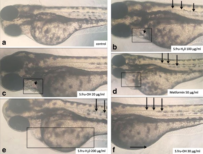
Fig. 1.
Embryos treated with different concentrations of S.fru-OH or S.fru-H2O at 96 hpf. a An example of a control (untreated) embryo with a normally developed straight spine without any visible signs of bleeding. b The short arrow points to a darkened region where a cyst is forming. The boxed zone highlights the pericardial region. c The short arrow points to a pink discolouration within the pericardial zone (shown using a rectangle). d A distinct pink sphere is visible within the pericardial zone (rectangle) and long arrows point to a curving spine. e A clear mass of tissue and an enlarged abdominal region is highlighted by the rectangle. f Long arrows point to a slightly curving spine. Pericardial cyst formation caused by exposure of zebrafish embryos to S.fru-H2O at 100 and 200 μg/ml was also accompanied by enlarged bellies (shown here by a horizontal arrow)

