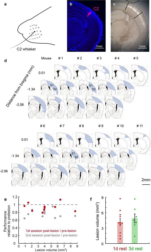
Extended Data Figure 4 |. Behavioral performance after lesions did not correlate with lesion size.
a, Mice performed the task with a single C2 whisker. b, The location of the C2 barrel in a coronal section. The C2 barrel was functionally mapped with intrinsic imaging and Alexa-conjugated cholera toxin subunit B (CTB, red) was injected into the center of the C2 barrel. Blue: DAPI. Mappings in b and c are used to inform the location of lesions made relative to the C2 barrel column. c, Equivalent location in section of an Nr5a1-eYFP animal with barrels fluorescently labeled in L4 (white) overlaid on bright-field image to show extent of barrel cortex relative to section (black lines). C2 was located ~1.2–1.5 mm posterior to bregma, varying slightly between animals. Lesions were centered around C2. d, Size and locations of contralateral barrel cortex lesions for the 11 mice with 1-day rest shown in Fig. 3 (arranged from largest to smallest by lesion volume). For each mouse, three locations along the anterior-posterior axis are shown overlaid on atlas images reproduced with permission from Paxinos & Franklin, 200142. In a few mice (e.g., mouse 1, 3, 8), lesions extended into the secondary somatosensory area (S2). Numbers along anteroposterior axis indicate approximate location relative to bregma. e, Lesion size did not correlate with the degree of impairment on the first (gray) or second post-lesion session when behavioral performance recovers (red). Performance was normalized to the pre-lesion performance for each animal. f, Lesion sizes were similar between groups with 1 or 3-days of rest after lesioning.

