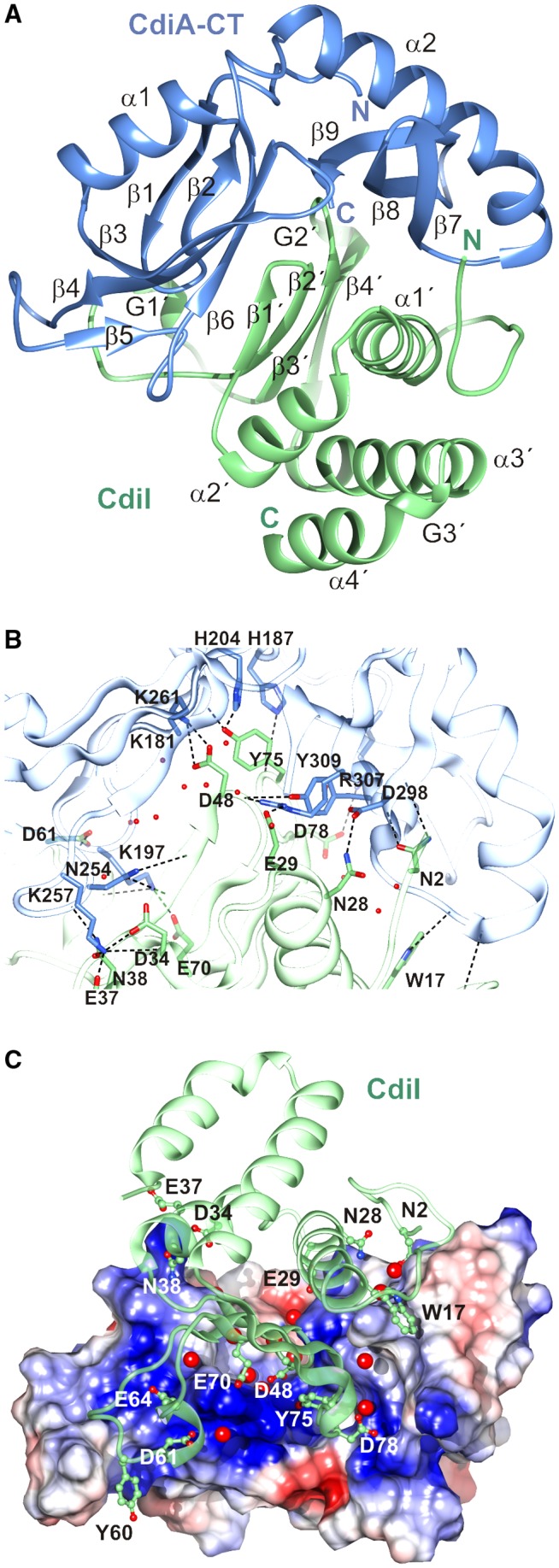
Figure 1.

Structure of the CdiA‐CT/CdiISTECO31 complex. A. The CdiA‐CT/CdiISTECO31 complex is depicted in cartoon with the toxin domain colored blue and the immunity protein colored green. Secondary structure elements are labeled with CdiISTECO31 elements denoted by a prime (′) symbol. B. The toxin‐immunity protein interface is depicted with selected side‐chains forming direct hydrogen bonds (black dashed lines) shown in a stick representation. Water molecules that mediate interactions are shown as red spheres. C. Charge complementarity at the toxin‐immunity protein interface. The electrostatic potential of the toxin surface was calculated using Coulomb's law with Chimera (Pettersen et al., 2004). Potentials range from –10 kcal/mol*e (red) to +10 kcal/mol*e (blue). Water molecules that mediate interactions are shown as red spheres. [Colour figure can be viewed at http://wileyonlinelibrary.com]
