Figure 1.
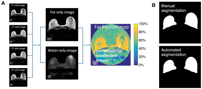
Derivation of the fat fraction map from data acquired using the radial IDEAL-GRASE technique and the breast segmentation results. A, (a)–(d) Images obtained from data acquired with each of the 4 gradients echoes of the radial IDEAL-GRASE sequence and the calculated (e) fat-only image, (f) water-only image and (g) fat fraction map (calculated voxel-wise as the ratio of the fat signal to the sum of the fat and water signals). B, the corresponding manual, and automated breast segmentation results. The average Dice index was 0.9121 with a standard deviation of 0.0306 based on the data from Sample 1.

