
Figure 1. Phosphatidylserine Is Dynamically Exposed on Degenerating Dendrites after Injury and during Dendrite Remodeling.
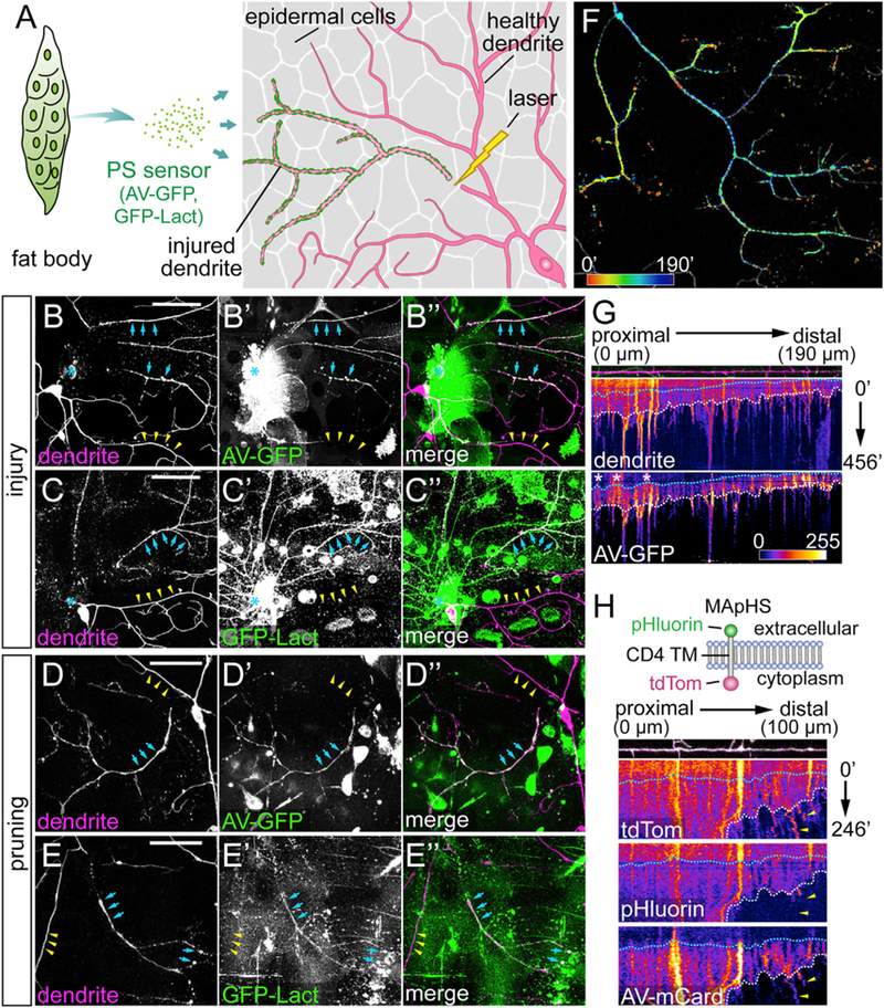
(A) Experimental design for visualizing PS exposure on injured dendrites of C4 da neurons.
(B–C”) Labeling of injured C4 da dendrites by AV-GFP (B–B”) and GFP-Lact (C–C”) at 4–5 hr after injury (AI). Blue arrows: injured dendrites; yellow arrowheads: uninjured dendrites; blue asterisk: injury sites.
(D–E”) Labeling of severed C4 da dendrites by AV-GFP (D–D”) and GFP-Lact (E–E”) during dendrite pruning between 6 and 7 hr after puparium formation (APF). Blue arrows: severed dendrites; yellow arrowheads: not-yet-severed dendrites. Some hemocytes are visible in the GFP channel in (B)–(D”) because Dcg-Gal4 also drives expression in hemocytes.
(F) Temporal sequence of AV-GFP binding to injured dendrites (Video S3). Red: early binding; blue: late binding. Indicated times are relative to the first frame.
(G) Kymographs of morphology and AV-GFP binding of an injured dendrite. The top panel shows the straightened dendrite with indicated distances relative to the proximal (left) end. The middle and bottom panels show kymographs of tdTom and AV-GFP signals, respectively, with indicated times relative to the first frame. White dotted lines: timing of dendrite fragmentation based on the continuity of the dendrite signal; blue dotted lines: timing of AV-GFP initial binding to the dendrite; white asterisks: interfering AV-GFP signals from injured dendrites of other da neurons.
(H) Kymographs of engulfment and AV-mCard binding of an injured dendrite. Top panel: diagram of the MApHS marker (adapted from Han et al., 2011). Dendrite and kymograph panels are arranged similar to (G). White dotted lines: timing of dendrite fragmentation; blue dotted lines: timing of AV-mCard initial binding; yellow arrowheads: engulfed dendrite debris (based on the loss of pHluorin) still associated with AV-mCard.
Scale bars: 50 μm (B–E). See also Figure S1 and Videos S1, S2, S3, and S4.