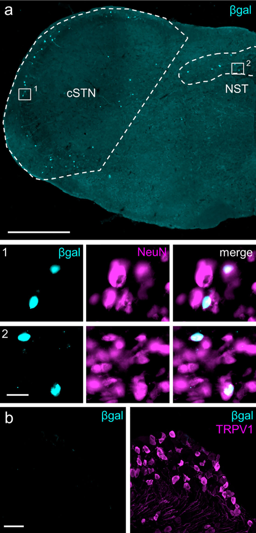
FIGURE 1.

Aromatase expression in the medulla and trigeminal ganglia (TG). (a) Representative section from aromatase reporter mouse illustrates nuclear β-galactosidase (βgal) expression in laminae I and V of the caudal spinal trigeminal nucleus (cSTN, area demarcated by dashed lines) and in the nucleus of the solitary tract (NST, area demarcated by dashed line). Co-staining for the neuronal marker, NeuN, shows complete overlap with β-gal, examples of which can be seen in insets 1 and 2. Image is stitched from single 20X images. Scale bar: 500 μm; insets: 20 μm. (b) No β-gal signal was detected in the TG (left panel). For comparison, right panel illustrates TRPV1- immunoreactive neurons in the same section. Scale bar: 50 μm. [Color figure can be viewed at wileyonlinelibrary.com]
