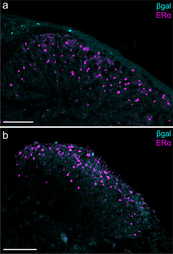
FIGURE 3.

Aromatase and estrogen receptor α (ERα) expression in the medullary and spinal dorsal horns. (a) Caudal spinal trigeminal nucleus: β-gal+ cells are visible in lamina I, whereas ERα-expressing cells are distributed throughout laminae I and II. Scale bar: 100 μm. (b) Sacral spinal cord: There is also no overlap of β-gal and ERα in the spinal cord. Scale bar: 100 μm. [Color figure can be viewed at wileyonlinelibrary.com]
