Figure 2.
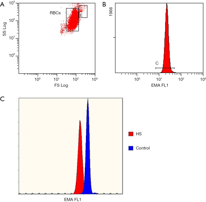
Eosin-5’-maleimide test. (A) Red blood cells are gated in gate “RBCs”, gate “E” contains doublets of erythrocytes. Primary gating is based on forward scatter vs. side scatter logarithmic scale; (B) EMA-bound cells’ fluorescence is measured in the first channel of fluorescence at 488 nm excitation wavelength and presented as mean fluorescence intensity; (C) the overlay histogram of fluorescence of EMA-stained RBCs from the HS subject (first, lower peak) and a healthy control (second peak), respectively. Peaks differ in mean fluorescence intensity.

