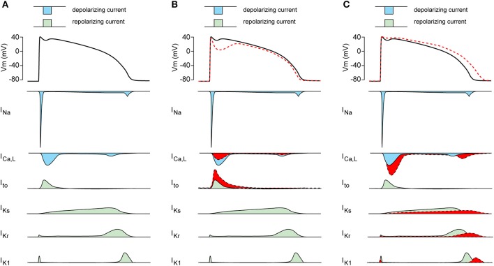
Figure 2.
(A) Schematic drawing of the cardiac action potential (AP) and its underlying membrane currents. INa, Na+ current; ICa,L, L-type Ca2+ current; Ito, transient outward K+ current; IKs, slow component of the delayed rectifier K+ current; IKr, rapid component of the delayed rectifier K+ current; IK1, inward rectifier K+ current. (B) Schematic drawing of AP plateau suppressing ion channels changes (in red). (C) Schematic drawing of AP prolonging ion channels changes (in red).

