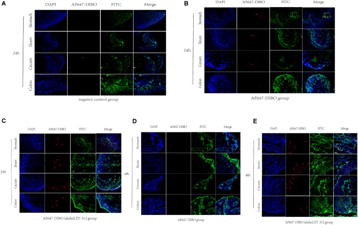
FIGURE 3.
Imaging of mice using confocal microscopy (A) Immunofluorescence of C57BL/6 mice’s stomach, ileum. Cecum and colon in negative control group. (B,C) Immunofluorescence of C57BL/6 mice’s tissue in AF647-DIBO group (B) and AF647-DIBO labeled ZY-312 group (C) for 24 h after treatment. (D,E) Immunofluorescence in AF647-DIBO group (D) and AF647-DIBO labeled ZY-312 group (E) for 48 h after treatment. Data are representative of at least three independent experiments.

