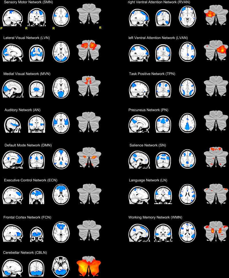
FIGURE 2.
Illustration of the 15 RSNs identified in this study. From top left: sensory motor network (SMN), lateral visual network (LVN), medial visual network (MVN), auditory network (AN), default mode network (DMN), executive control network (ECN), frontal cortex network (FCN), right (R) and left (L) ventral attention networks (VAN), task positive network (TPN), precuneus network (PN), salience network (SN), language network (LN), working memory network (WMN) and the cerebellar network (CBLN). Each RSN is presented as a blue mask on a sagittal, coronal and axial view. For each RSN, the last column of each trio of views show the cerebellar areas (in red-yellow scale) of the network plotted on a flatmap of the cerebellar cortex. With the exception of CBLN which involved the almost the entire cerebellum, nine RSNs showed at least one cerebellar node.

