. 2018 Oct 8;113(10):e180311. doi: 10.1590/0074-02760180311

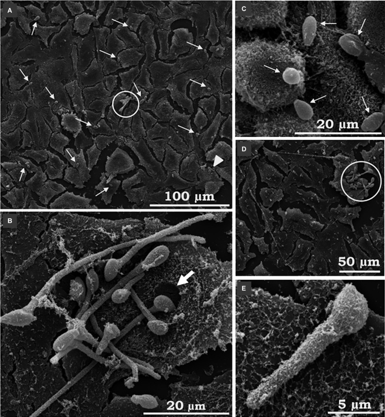
Fig. 3: scanning electron microscopy (SEM) of the interaction between Scedosporium apiospermum conidia and A549 cells. The images revealed many conidial cells adhered to animal cells and some germinated conidia (A-E). Thin white arrows indicate the associated conidia; the white arrowhead shows dispersed germinated conidia, while the open circles show “nests” of germinated conidia. The thick white arrow points to a hollow corresponding to the exact location where the conidium was anchored to the mammalian cell (B). A filamentous network produced by epithelial cells can be seen surrounding germinated conidia (E). The micrographs are representative images of three independent experimental sets.

Fig. 3: