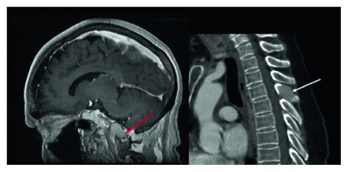
Figure 1.

Magnetic resonance imaging (MRI) of the head and spine. MRI captured a 2.8 × 2.2 × 1.9-centimeter enhancing lytic mass centered in the left clivus and occipital condyle (red arrow). Additionally, an expansile soft tissue lesion was noted in the T4 spinous process (white arrow).
