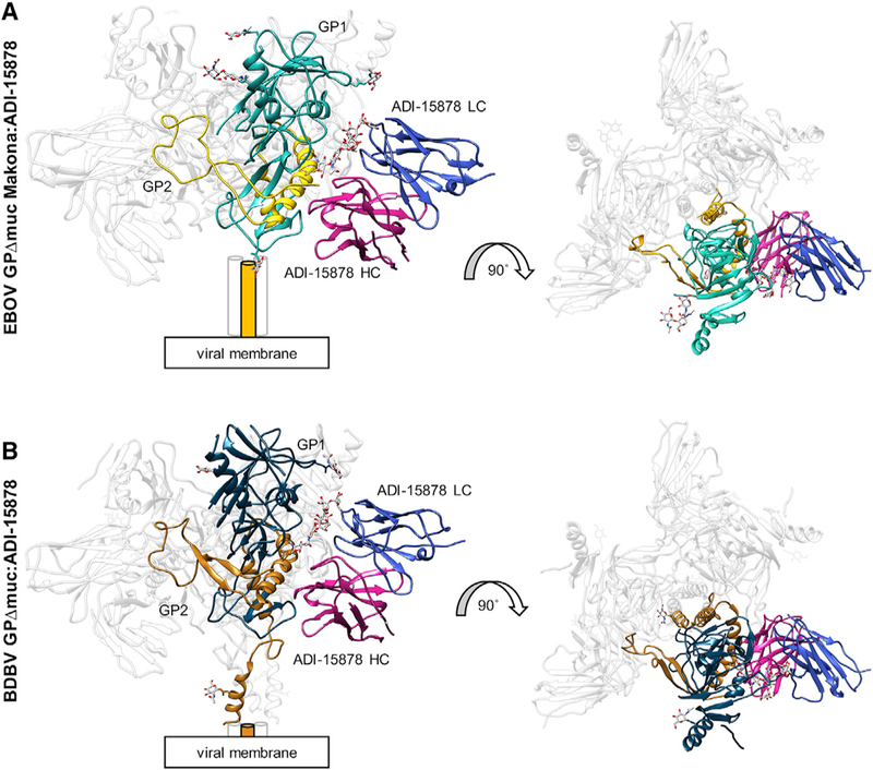
Figure 3. Cryo-EM Structures of ADI-15878 Fab Bound to EBOV/Mak GPΔmuc and BDBV GPΔmuc.

(A) Model of EBOV/Mak GPΔmuc bound to ADI- 15878 Fab. Shown are side (left) and top (right) views. We built four glycans in GP1 (cyan) and one in GP2 (yellow) and only the variable (Fv) domain of ADI-15878, with the light chain (LC) in blue and the heavy chain (HC) in magenta. Cylinders represent the HR2 domain, which were not built in this model. (B)Model of BDBV GPΔmuc bound to ADI-15878 Fab. We describe the core GP structure in Figure 2 and only built the ADI-15878 Fv domain, as described in (A).
See also Figures S1 and S2.
