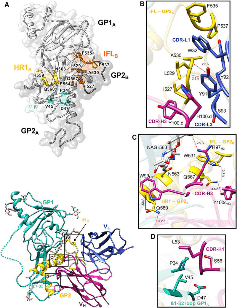
Figure 4. Details of the ADI-15878 Epitope in EBOV/Mak GPΔmuc.

(A) The ADI-15878 epitope footprint, which spans two GP1/2 protomers, including the β1-β2 loop in GP1A (cyan), HR1 in GP1A (yellow), and the IFL in GP2B (orange). All residueswithin 4Å of ADI-15878 in our model are labeled and highlighted.
(B) Details of the ADI-15878 epitope at the IFL in GP2B. A loose hydrophobic network is formed between Y100cH3, H100dH3, Y91L3, Y92L3, W32L1, and IFL residues 527–530. The peptide backbone O from Y92L3 is also within hydrogen-bonding distance ofthe backbone N from L529 and A30 on the IFL. W32L1 inserts itself between F535 and L529 of the IFL.
(C) Details of the ADI-15878 epitope at HR1 in GP2A. W99H3 stacks against Q560HR1 at a distance of ~3.8 Å, similarto the interaction ofW531IFL with Q567. Y100cH3 and R97H3 are within hydrogen bonding distance of Q567 and NAG-563HR1, respectively. Also, NE2 of Q560 is within hydrogen bonding distance of the main chain O of S30H1.
(D) Residues L54H1 and S56H1 are within 4 Å of portions of the β1-β2 loop in GP1A and may mediate contacts. Dashed lines represent regions that we did not build in our model but were included in our construct.
See also Figures S2, S3, and S4.
