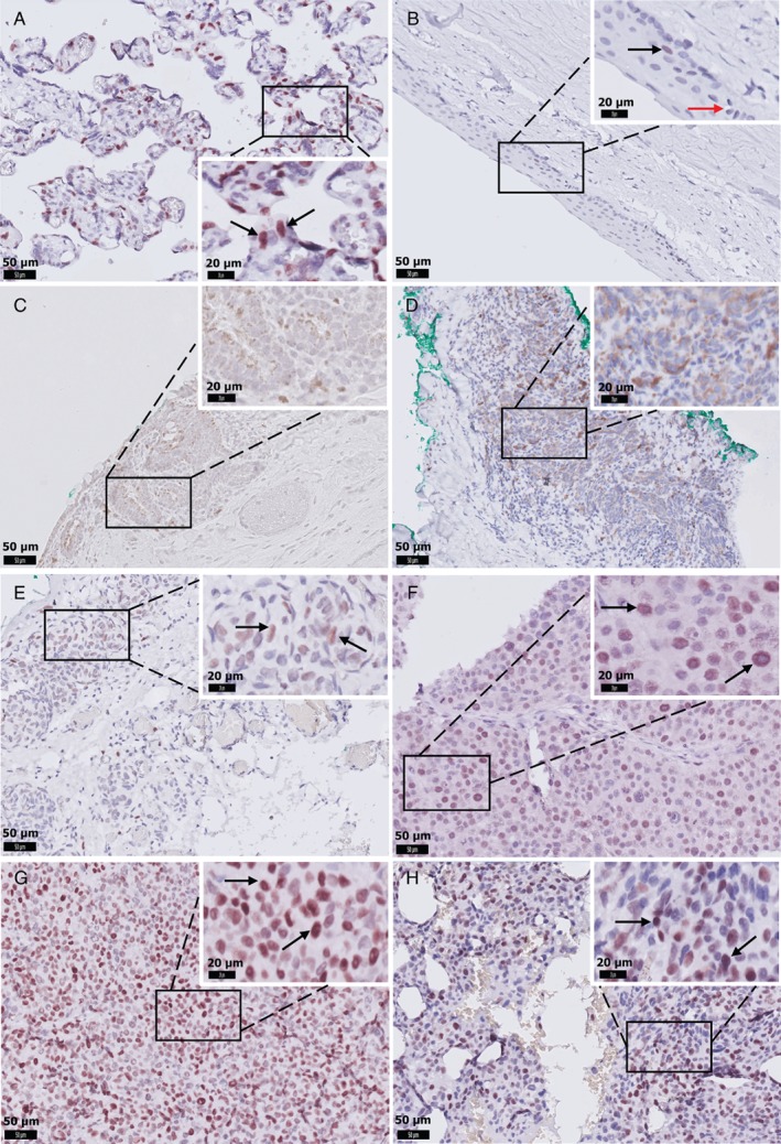
Figure 1.

Representative images of EZH2 IHC staining in CM. (A) Positive control staining of placental tissue. (B) EZH2‐positive keratinocytes (black arrow), and EZH2‐negative melanocytes in the normal conjunctiva (red arrow). (C) EZH2 is absent in primary acquired melanosis. (D) CM with negative nuclear staining in all tumour cells. (E) CM with weak, (F) moderate, or (G) strong nuclear staining in more than 50% of tumour cells. (H) Strong nuclear staining in tumour cells in a lymph node metastasis. D and E were considered EZH2‐low expression (scored ≤3); F, G, and H were scored >3 and considered EZH2‐high expression. Scale bars are 50 μm (original images) and 20 μm (insets). Black arrows indicate EZH2‐positive cells.
