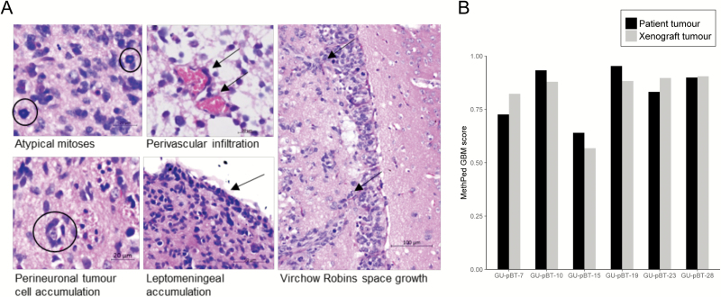
Figure 3.
Histology and methylation-based classification confirms GBM identity of xenograft tumours. (A) Enlargement of classical GBM growth pattern with asymmetric cell division; ring mitosis, structure of Scherer; axonal cell growth, perivascular infiltration and lepto-meningal accumulation of tumour cells. (B) MethPed classified the xenotransplanted tumours as GBM, with similar scores as for the patient tumours.

