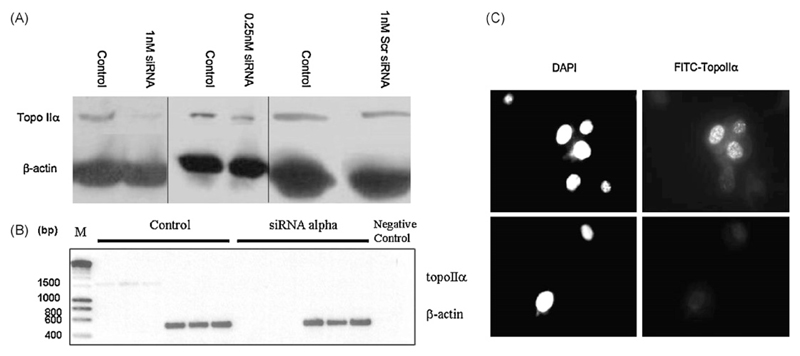
Fig. 1.
Western blots of topoisomerase IIα and β-actin in hTERT-RPE cells incubated for 12 h with 1 nM and 0.25 nM siRNA against topo IIα or 1 nM scrambled (Scr) siRNA (A). (B) RT-PCR products (reactions performed in triplicate) from cDNA extracted from control hTERT-RPE cells, and cells treated with siRNA against topo IIα (1 nM, 12 h). Negative control represents an RT-PCR reaction without cDNA and M stand for marker. (C) Immunocytochemistry with hTERT-RPE cells incubated with or without 1 nM siRNA against topo IIα for 12 h using primary anti-topo IIα antibody, and FITC-labelled secondary antibody (right panels). Nuclei (left panels) are stained with DAPI (magnification 63×).

