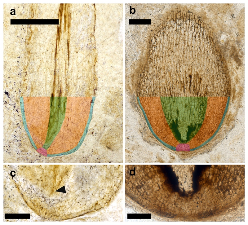
Extended Data Figure 3. Fundamental tissues present in a rooting axis apex preserved after growth had finished (a) and a rooting axis meristem preserved during active growth (b).
a, b, Root apices with fundamental tissue types colour coded (blue, epidermis; pink, promeristem; orange, cortex; and green, procambium). c, d, Magnified images of the apical regions of (a) and (b), respectively. The presence of differentiated vascular tissue (arrowhead c) close to the tip of the apex indicates this apex was not active at the time of preservation. By contrast, in (d) there is no differentiated vascular tissue. Instead the apex is characterised by large numbers of cells and cell size gradually increases with distance from the tip indicating the apex was active when fossilised. (a, c) NHMUK V.15642 – same specimen as illustrated in (Fig. 1a), (b, c) OXF 108 – same specimen as illustrated in (Fig. 1d; 2a, b). Scale bars (a) 500 µm, (b) 250 µm, (c) 150 µm, (d) 100 µm.

