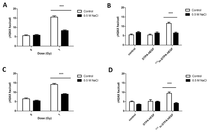
Fig. 5.
(A, B) MDA-MB-468 and (C, D) 231-H2N cells were incubated with hypertonic medium, exposed to (A, C) IR, and stained for γH2AX after 30 minutes. (B, D) After hypertonic treatment, cells were incubated with 111In-DTPA-hEGF (18 MBq/μg; 12 nM) or DTPA-hEGF (12 nM) and stained for γH2AX immediately.

